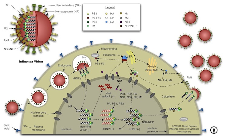
چرخه تکثیر ویروس آنفلوآنزا
+ نوشته شده در جمعه ۱۳۸۷/۰۱/۳۰ ساعت توسط m.ghane
آنفلوانزاي پرندگان H5N1 از طرق مختلفي همچون پرنده به پرنده، پرنده به انسان، محيط به انسان و شايد از انسان به انسان مي تواند انتقال يابد. 
انتقال از پرنده به پرنده :
پرندگان آبزي بخصوص اردك هاي وحشي وقتي به آنفلوانزاي پرندگان آلوده شوند، علائم بيماري در آنها ظاهر نميشود و اين پرندگان فقط ناقل سالم مي باشند و بطور كلي اردك هاي وحشي مخازن طبيعي اين ويروس در طبيعت ميباشند و بدون اينكه به بيماري شديدي مبتلا شوند قادرند ويروس را در محيط اطراف خود منتشر كنند ولي مرغ و خروس ها شديداً مستعد ابتلا به بيماري آنفلوانزاي پرندگان هستند. از آنجا كه ويروس در ترشحات بدن خصوصاً بزاق، ترشحات بيني و مدفوع پرندگان به مقادير فراوان موجود است، بيماري براحتي در جريان تماس مستقيم پرندگان وحشي با پرندگان اهلي و از طريق آب آشاميدني مشترك باعث آلودگي پرندگاني مثل مرغ و خروس مي شود. با آلوده شدن مرغ و خروس ها بيماري به آساني از يك مرغداري به مرغداري ديگر انتقال مي يابد زيرا تعداد زيادي ويروس در فضله پرندگان وجود دارد و باعث آلودگي گرد و غبار و خاك مي گردد و ضمناً ويروس از طريق هوا به آساني از پرنده اي به پرنده ديگر منتقل مي شود . لوازم و اشياء آلوده، غذاها، قفسه ها، لباس ها و بويژه كفش هاي كارگران مرغداري، قفسها و كاميون هاي مخصوص حمل پرندگان نيز موجب انتقال ويروس از محلي به محل ديگر مي شوند. ويروس همچنين از طريق پاها و بدن حيواناتي نظير جوندگان كه نقش ناقل مكانيكي را ايفا مي نمايند نيز ممكن است منتقل گردند.

مقدمه :
در حالي كه كشورهاي پيشرفته جهان در طي چند دهه اخير مشكل بيماريهاي واگيردار را تا حد زيادي حل كرده و تمامي توان و امكانات خود را براي مبارزه با بيماريهاي غير واگير منجمله بيمارهاي قلبي – عروقي و سرطانها متمركز كرده بودند ، ناگهان در سال 1981 ميلادي (1360 شمسي ) نوعي بيماري عفوني در دنيا پيدا شد كه به سرعت درهمه نقاط جهان گسترش پيدا كرد و اميد به ريشه كني بيماريهاي واگيردار را حتي در كشورهاي پيشرفته تبديل به ياس نمود. اين بيماري كه بصورت يك مشكل عمده بهداشتي خود را نشان داد ايدز نام دارد . در اين بيماري دستگاه دفاعي بدن شديداًدچار اختلال مي شود.
As previously mentioned, Gram-positive bacteria are characterized by their blue-violet color reaction in the Gram-staining procedure . The blue-violet color reaction is caused by crystal-violet, the primary Gram-stain dye, complexing with the iodine mordant. When the decolorizer is applied, a slow dehydration of the crystal-violet/iodine complex is observed due to the closing of pores running through the cell wall. Because the crystal-violet is still present in the cell, the counter stain is not incorporated, thus maintaining the cell's blue-violet color. If you recall, most cell walls contain peptidoglycan, a molecule made of amino acids and sugar. A distinguishing factor among Gram-positive bacteria is that roughly 90% of their cell wall is comprised of peptidoglycan and a Gram-positve bacteria can have more than 20 layers of peptidoglycan stacked together to form the cell wall. That's pretty thick! Examples of common Gram-positive cells are Staphylococcus aureus and Streptococcus cremoris, a bacterium used in dairy production.
The Gram-positive cocci are grouped together based on their Gram-stain reaction, thick cell wall composition, and spherical shape. Most of the organisms in these groups are members of the Micrococcaceae family All of the organisms in these groups are non-endospore forming chemosynthetic heterotrophs. We will discuss only the clinically relevent bacteria from Micrococcus and Staphylococcus of the Micrococcaceae family. Streptococcus and Enterococcus(formerly a species of Streptococcus) are discussed as well because of the many diseases they inflict on humans. The chart below shows the paths to identification of the genera discussed.
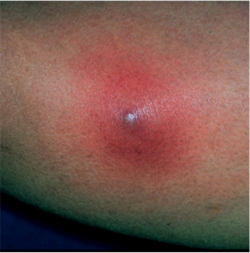
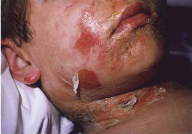
Clinically, the most important genus of the Micrococcaceae family is Staphylococcus. The Staphylococcus genus is classified into two major groups: aureus and non-aureus. S. aureus is a leading cause of soft tissue infections, as well as toxic shock syndrome (TSS) and scalded skin syndrome. It can be distinguished from other species of Staph by a positive result in a coagulase test(all other species are negative).
The pathogenic effects of Staph are mainly asssociated with the toxins it produces. Most of these toxins are produced in the stationary phase of the bacterial growth curve. In fact, it is not uncommon for an infected site to contain no viable Staph cells. The S. aureus enterotoxin causes quick onset food poisoning which can lead to cramps and severe vomiting. Infection can be traced to contaminated meats which have not been fully cooked. These microbes also secrete leukocidin, a toxin which destroys white blood cells and leads to the formation of pus and acne. Particularly, S. aureus has been found to be the causative agent in such ailments as pneumonia, meningitis, boils, arthritis, and osteomyelitis (chronic bone infection). Most S. aureus are penicillin resistant, but vancomycin and nafcillin are known to be effective against most strains.
Microcoocus is a Gram-positive, aerobic bacterium which is a member of the Micrococcaceae family. Micrococcus cells can be observed under the microscope as spherical cells forming pairs or clusters. If cultured in broth or on nutrient agar, the colonies may be red or yellow when observed unstained. Although these bacteria are a common human skin contaminant, they are relatively harmless to humans because they maintain a saprophytic lifestyle. They can also be found in freshwater environments or in soil. Three common species of Micrococcus are M. luteus, M. roseus, and M. varians.